The BTH Before & After Library

Gingival Masks Before and After: 6 Clear Comparisons

Gingival masks can restore missing gum contour without surgery or permanent alteration of the teeth. The next question is how well it can be done.

This page brings together six sourced gingival mask before-and-after cases so patients can see the range of results in one place. You may also see these appliances called gingival veneers, gingival epitheses, or removable gingival prostheses. The examples include published clinical cases and commercial work, with close-up images chosen to make the treatment easier to understand without requiring readers to dig through technical papers, scattered websites, or difficult clinical photography.

The goal is simple: make the option visible.

For patients who have spent months or years searching for answers, clear examples matter. They show what gingival masks can cover, how natural they can look, how much tissue can be replaced, and where the differences between one result and another become apparent.

This is not a promise of what every patient can achieve. It is a clearer view of what has already been done.

Sourced clinical examples

Ultimate Gingival Masks and Gingival Veneers Collection

Use the sliders to compare each documented result. Photographs differ in crop, lighting, angle, resolution, and timing, so BTH does not assign clinical scores from images alone. We do identify visible features patients and professionals should discuss.

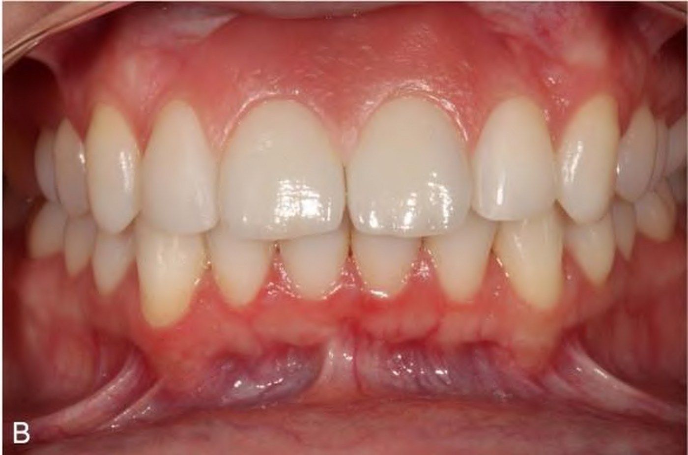
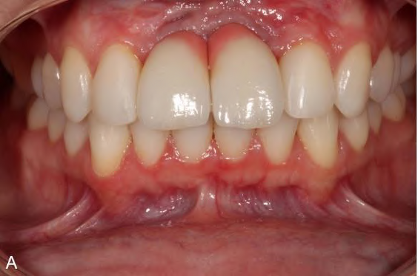
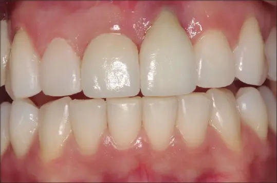
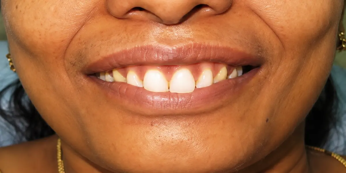
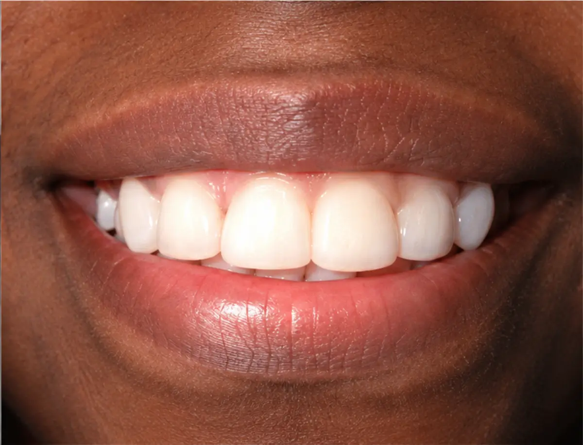
Patient smiling with the acrylic gingival veneer documented by Adhershitha and Anilkumar The same patient smiling before placement of the acrylic gingival veneer BEFORE WITH VENEER
CASE 01 · PUBLISHED CASE REPORT

Adhershitha and Anilkumar

An acrylic gingival veneer restored the visible gingival contour across the anterior smile without surgery or permanent alteration of the teeth.

BTH visual note: the full-smile effect.

The paired photographs show why these appliances must be judged in the smile, not only in an isolated close-up. Replacing the missing pink field changes the entire visual balance.

Clinical photographs: Adhershitha & Anilkumar, 2023. Image enhanced for clarity. Read the case report.
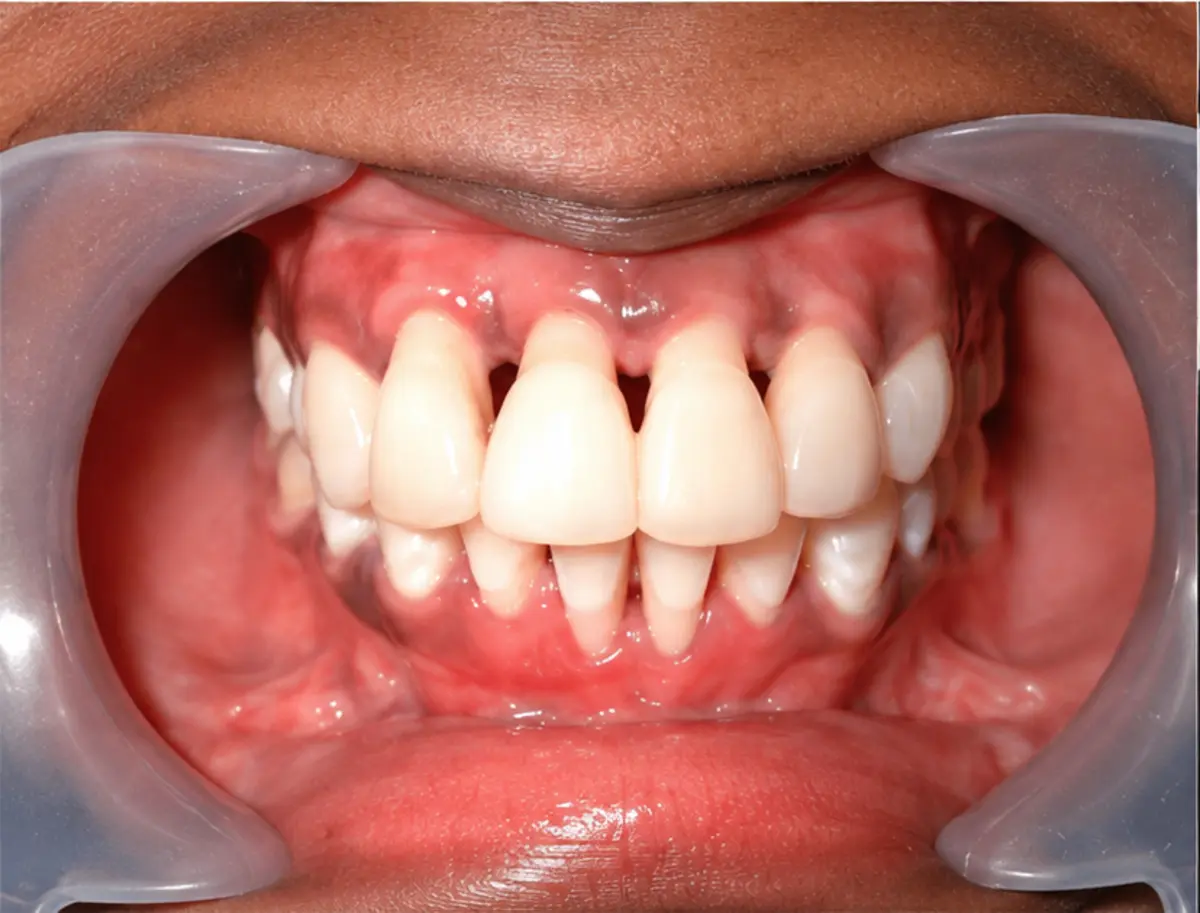
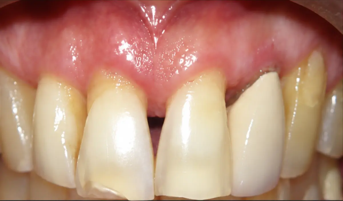
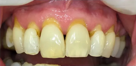
Patient smiling with the acrylic gingival mask documented by Antony and Khan The same patient before the gingival mask, showing severe recession and open interdental spaces BEFORE WITH MASK
CASE 02 · PUBLISHED CASE REPORT

Antony and Khan

This acrylic gingival mask concealed severe recession and open interdental spaces after periodontal treatment.

BTH patient-outcome note: confidence returned.

At two-year follow-up, the authors reported comfortable continued use, virtually no problems, and renewed confidence facing other people.

Source: Antony & Khan, 2013. Read the paper. · View the figure.
Smile with the gingival epithesis made by Diana Ankli and colleagues The same smile without the gingival epithesis WITHOUT WITH MASK
CASE 03 · PUBLISHED REPORT

Diana Ankli and colleagues

A removable epithesis produced in Belo Horizonte. The finished appliance restores the papillary spaces and broader gingival field across the visible anterior region.

BTH visual note: the benchmark.

The close color integration is the immediate strength. The mask reads as gingiva before it reads as an appliance. Contour and surface variation keep the pink field from looking flat or generically molded.

Source: Diana Ankli et al. Images cropped for comparison; clinical content unchanged. Read the paper.
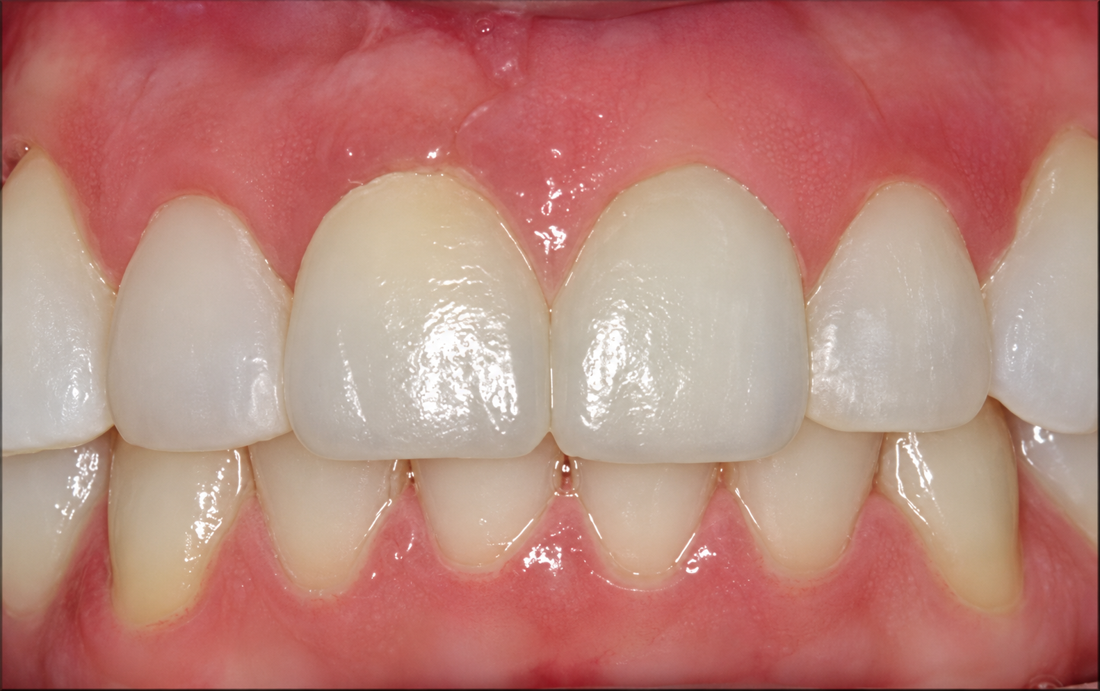
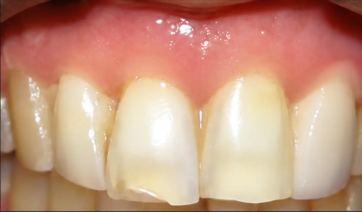
Enhanced clinical view with the gingival veneer covering recession associated with a malpositioned anterior implant The same patient before placement of the gingival veneer, showing peri-implant recession BEFOREWITH VENEER
CASE 04 · PUBLISHED IMPLANT CASE

Viviane Ankli and colleagues

This removable gingival veneer was made to cover peri-implant gingival recession associated with a malpositioned single implant in the upper front teeth. Move the divider to compare the untreated recession with the completed veneer.

BTH visual note: fantastic work.

The veneer is localized to a difficult defect yet blends into the surrounding gingiva with convincing color, contour, and stability. It shows how a removable solution can preserve the implant while restoring the missing pink architecture.

Clinical photographs from Gingival Veneer Used as Prosthetic Solution for Esthetic-Compromised Malpositioned Dental Implant — Viviane Ankli, Francisco Ivison Rodrigues Limeira, Monica Yamauti, and Tassiana Cançado Melo Sá. Images cropped for comparison; the veneer image was enhanced for clarity. Read the paper.
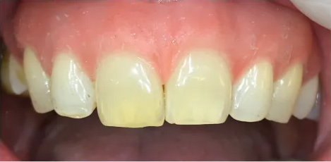
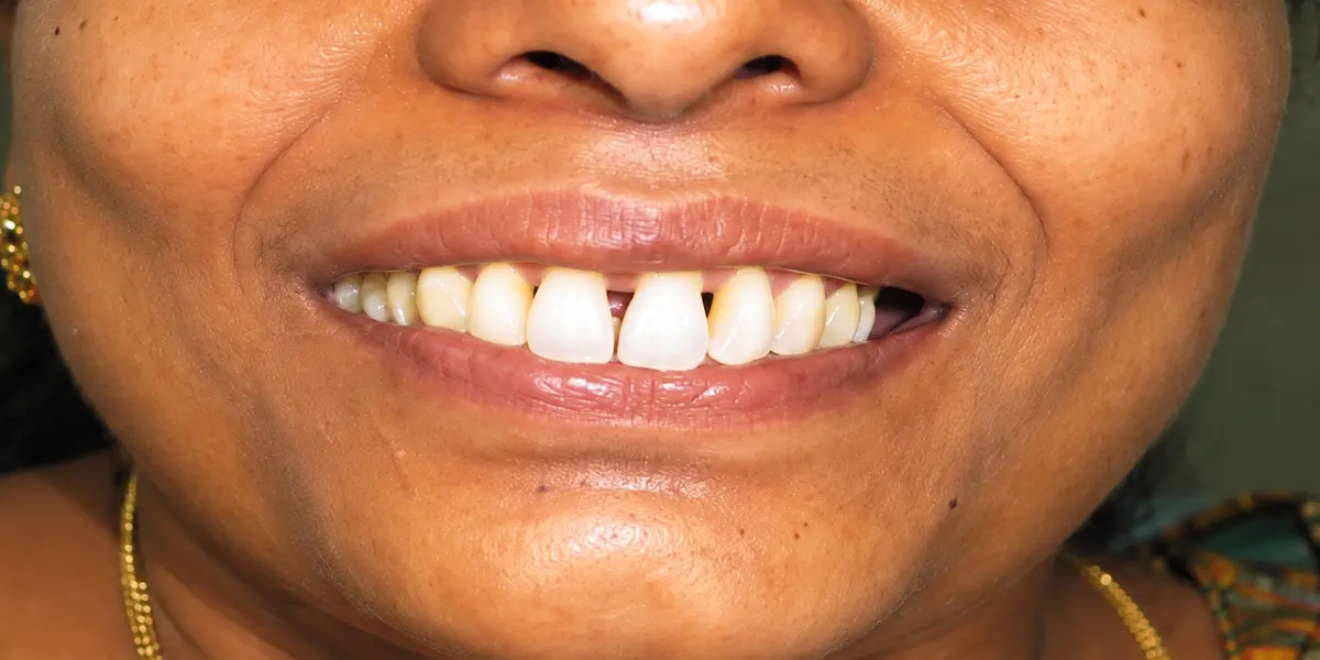
Smile after placement of the gingival veneer documented by Thakkar and colleagues The same smile before placement of the gingival veneer BEFORE WITH VENEER
CASE 05 · PUBLISHED CASE SERIES

Thakkar and colleagues

This published case shows a gingival veneer restoring the visible gum contour across multiple areas of recession and open gingival embrasures.

BTH visual note: coverage across a broad defect.

The veneer closes the central black triangle and replaces a wider field of missing tissue. The paired view makes both the extent of the original defect and the change in visible contour immediately understandable.

Clinical photographs from Gingival Veneers: A Case Series for Treatment of Multiple Gingival Recession and Black Triangles — Priya Vijay Thakkar, Amol Beldar, Supriya Murkute, and Mitali Thamke. Images cropped for comparison; clinical content unchanged.
Upper front teeth with an e-Dentic gingival epithesis The same upper front teeth without the gingival epithesis WITHOUT WITH EPITHESIS
CASE 06 · LABORATORY EXAMPLE

e‑Dentic

The Swiss laboratory describes very thin, flexible silicone or durable resin options intended to provide a natural gingival appearance and patient comfort.

BTH visual note: restoration with room to scrutinize.

The after image restores substantial volume and covers exposed root areas. It also shows why patients need close-up examples: shade, opacity, papilla shape, and the upper edge all affect whether the result blends or announces itself.

Source: e‑DENTIC.CH. Original paired image separated into two crops for the slider. View the source page.
The BTH quality standard

“Pink” is not a color match.

Natural gingiva varies between people, within the same mouth, and across age and location. A serious prosthesis must be matched to the patient—not pulled from a generic idea of healthy gum color.

01

Hue, value, and chroma

Match the actual tissue under controlled viewing conditions. Too bright, too pale, too purple, or too saturated can expose the appliance immediately.

02

Translucency and depth

Living tissue is not an opaque slab. Layering, internal characterization, and controlled translucency can create depth instead of flat pink.

03

Contour and papillae

Replacement volume should follow the individual anatomy and meet each tooth convincingly—not simply bridge every opening with the same shape.

04

Surface texture

Subtle stippling and regional variation matter. Texture should support realism without becoming coarse, artificial, or difficult to clean.

05

Edge transition

The appliance margin should be thin, stable, comfortable, and as visually quiet as the anatomy allows—especially near a visible smile line.

06

Color over time

Material, diet, cleaning, and surface finish affect staining and stability. The first-day match is only the beginning of the result.

Diana Ankli’s result points toward the standard: the best mask is not the pinkest one. It is the one your eye stops noticing.
What the literature gives us

Turn scattered technique into shared knowledge.

BTH will keep extracting useful, source-linked details from case reports and materials research. These are discussion points for trained clinicians and technicians—not instructions for unsupervised self-treatment.

Record the patient’s tissue—not “standard pink.”

Research on natural gingiva documents meaningful differences by location and age. Modern shade research likewise tests whether commercial guides actually cover the colors found in real patients.

Sources: Ibusuki, 1975 · Gingival shade-guide compatibility

Build color in layers.

Prosthetic literature uses base color, localized characterization, vascular detail, and differing opacity to reproduce living tissue. The principle matters even when the exact material or appliance changes.

Source: Individualized gingiva-shade characterization

Material choice changes the tradeoffs.

Flexible silicone can produce a light, retentive appliance, while harder materials may offer different surface and color-stability behavior. The literature does not support treating all “gingival masks” as one interchangeable product.

Sources: Silicone gingival prosthesis case · Epithesis material comparison

Photograph the result honestly.

Useful records need the same angle, crop, scale, exposure, and lighting before and after. Include close views of margins and papillae. A flattering hero image cannot substitute for documentation.

BTH documentation standard for future submitted cases.

Record function with appearance.

Comfort, speech, saliva behavior, retention, removal, cleaning, eating, tissue response, and adjustments belong beside the photographs. A visually convincing mask can still fail the patient.

BTH patient-outcome standard informed by the functional concerns reported across gingival-prosthesis cases.
Proof removes one excuse

It exists. Now answer the patient.

A sourced result does not guarantee candidacy or outcome. It does establish that the conversation cannot end at “that isn’t a thing.” The responsible next answer is evaluation, fabrication coordination, or a referral to someone prepared to provide it.

See where access breaks →

Help build the definitive record.

Patients, clinicians, technicians, and laboratories: send us sourced cases, fabrication details, follow-up information, and results worth learning from. BTH will preserve the source, separate fact from observation, and make the useful information easier to find.

Submit a case or source